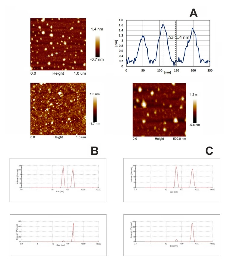
Figure 2.
Atomic force microscopy (AFM) topography and surface profile of fullerenol. C60(OH)36 was dissolved in water and precipitated on mica substrate (A). Dynamic light scattering (DLS) size distribution of the C60(OH)36 in water (B) and in 0.02 M phosphate buffer pH 7.4 (C), showing the intensity and volume distributions of fullerenol-water and fullerenol-PBS preparations. Intensity-weighted average values were used to determine the hydrodynamic size, while volume distribution data was used to determine relative amounts of nanoparticles (NPs). Graphs in Figure 2B,C were produced by the Zetasizer Nano-ZS software.

