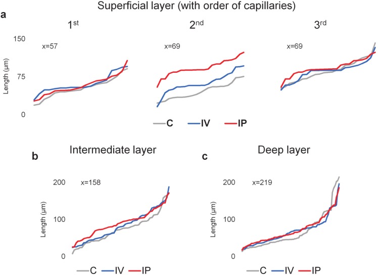
Figure 5.
Change in the distribution of capillary branch lengths (visualized in increasing order of length within each category) in the mouse retinal vasculature after imatinib IV and IP treatments in the (a) SL measuring the 1st-, 2nd-, and 3rd-order capillaries (determined by the branching points after arterioles) and on capillary branches in the (b) IL and (c) DL. n = 6 mice; x= number of measurements. The X-axis represents individual length measurements in ascending order.

