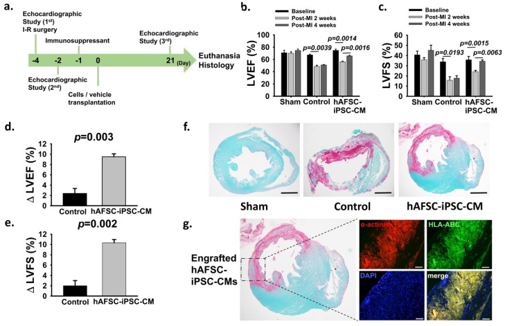
Figure 4.
Therapeutic effects of hAFSC-iPSC-CM transplantation. (a) Timeline for the efficacy study in which immunocompetent 8-week-old Sprague Dawley (SD) rats (350~400 g, both genders) received 60-minute ischemia, followed by reperfusion in the mid-left anterior descending coronary artery. Echocardiographic studies were performed prior to the infarct surgery 2 days prior to cell transplantation and 3 weeks after transplantation. (b,c) All the animals (including the control group and the hAFSC-iPSC-CM-treated group) had deteriorating cardiac function after myocardial infarction. Cardiac function studies (b, LVEF; c, LVFS) showed a significant improvement after hAFSC-iPSC-CMs treatment. (d,e) Changes in LVEF (∆ LVEF, d) and LVFS (∆ FS, e) from 2 days after myocardial infarction to 3 weeks after treatment were significantly greater in hAFSC-iPSC-CM-treated group. (f) A representative stain of a short-axis cross-section of SD rat hearts with picrosirius red and fast green. The fibrotic infarct region is red and healthy myocardium is green. There was no fibrotic tissue in the sham group. However, the infarct is transmural in the hearts of the control and the hAFSC-iPSC-CM-treated groups. Scale bar, 2mm. (g) hAFSC-iPSC-CM-engrafted heart demonstrating several human myocardium islands of considerable size in the infarcted region. These human myocardia were stained with human specific HLA-ABC antibody. Confocal immunofluorescence scale bar, 50 µm.

