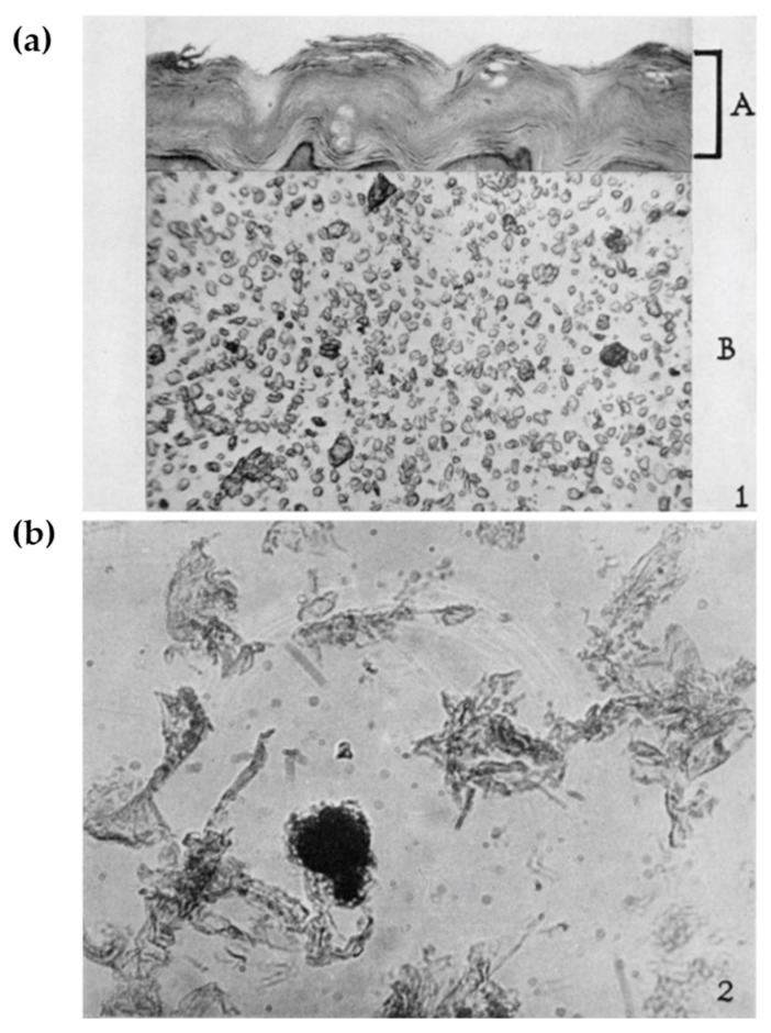
Figure 1.
(a) A cross-section of human plantar skin showing thickly cornified epithelium (A section). Pulverized callus showing single corneocytes. (b) Isolated cell-membrane-like remnants of corneocytes after exhaustive hydrolysis. Adapted from Ref. [13] (Copyright, 1955, by The Rockefeller Institute for Medical Research).

