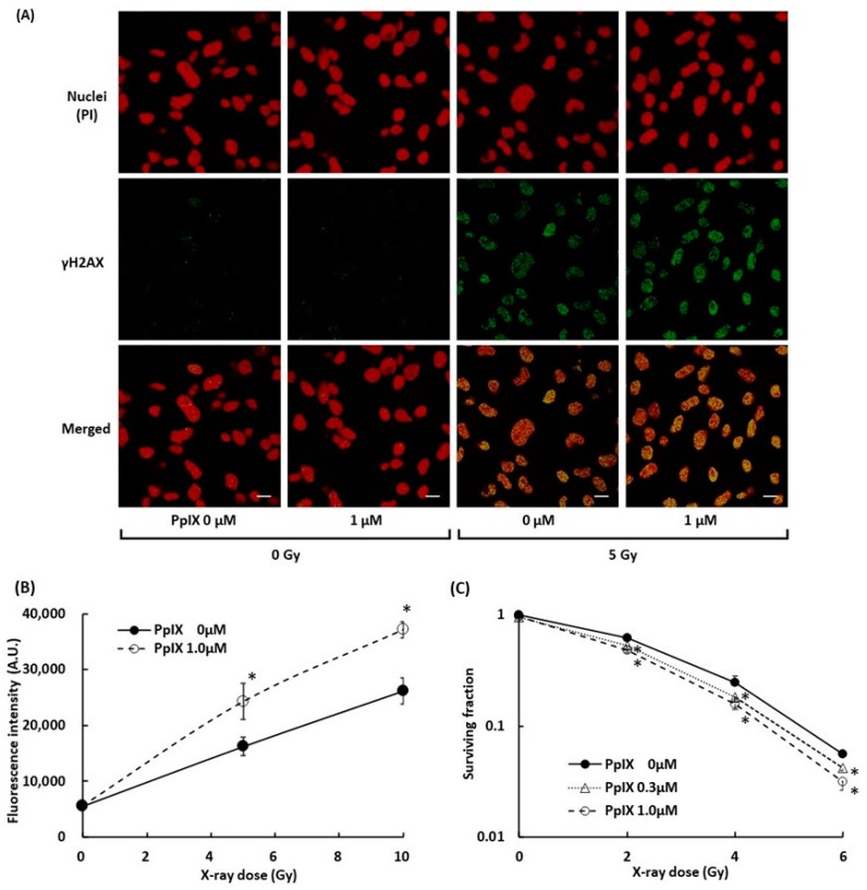
Figure 5.
Evaluation of double-strand breaks (DSBs) within nuclei by X-ray irradiation and enhancement by PpIX. (A) Fluorescence in cell culture was imaged using laser confocal microscopy. Subcellular localization of γH2AX (green) and propidium iodide (PI)-stained nuclei (red) in cells with and without exposure to 1 µM PpIX or 5 Gy X-ray radiation. Scale bars: 20 µm. (B) The fluorescence intensity of γH2AX. (C) The WST-8 cell viability assay for cellular responses to PpIX treatment and X-ray irradiation. Data are the means ± SD (n = 4). Statistical significance (p < 0.01) relative to the experiment performed without PpIX at the same irradiation dose indicated by (*).

