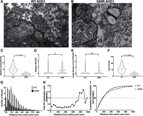
Figure 3.
Presynaptic infusion of G85R-SOD1 inhibited synaptic vesicle (SV) availability. Representative EM images illustrate morphology of AZs (labeled with red *) and numbers of SVs in fixed synapses infused with WT-SOD1 (A, n = 3) and G85R-SOD1 (B, n = 3). G85R-SOD1-infused synapses showed vacant AZs and occasional abnormal membranous structures (indicated by blue # and purple ⋀). C–F, Quantification of averaged vesicle number across all AZs from 102 WT- and 102 G85R-SOD1-YFP-infused synapses showed statistically significant reductions in the total vesicle number and in the electron lucid vesicle number by G85R-SOD1-YFP as compared with WT-SOD1. The clathrin-coated vesicles and the large electron lucid vesicles were comparable between WT- and G85R-SOD1-YFP-infused synapses. Nested one-way ANOVA was performed to compare WT and G85R-infused synapses (p < 0.0001) as well as across biological triplicates within each group (p > 0.05) ns: not significant. G, Averaged number of SVs per AZ were plotted along the distance from AZ (binned by 50 nm). H, Distance distribution plot of SVs in each 50-μm bin showed a global reduction of SVs from G85R-SOD1-YFP-infused synapses regardless of the distance from the AZs. Because of the drastically reduced numbers of SVs far (>675 nM) from the AZs in both WT and G85R synapses, the reduction seemed to disappear or even be reversed, however, the differences far from the AZ may not be significant due to the substantially decreased numbers of vesicles in that area for both WT and G85R synapses. I, Cumulative SV distribution plots showed similar distribution patterns of vesicles in WT (solid line) and G85R (dashed line) synapses, confirming the even inhibition of SV availability by G85R-SOD1 independent of the distance from the AZs.

