Figure 1.
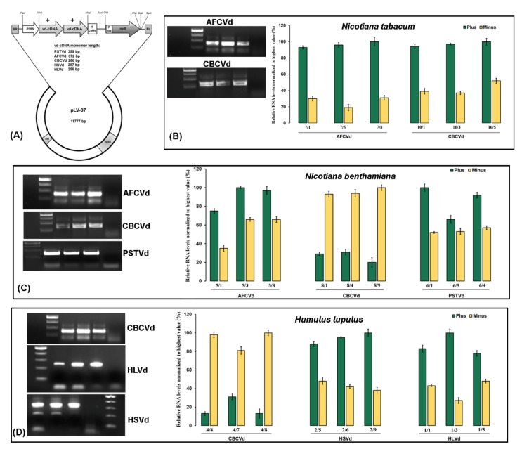
Schematic representation of dimeric infectious constructs, detection and quantification of viroids. (A) Schematic diagram of a plasmid containing the shown viroid (+) dimer created by cDNA cloning in SacI restriction site. The viroid (+) dimer was re-cloned from pPCR-Script to XhoI–XbaI sites of intermediary vector pLV-68. The final modified binary expression cassette harboring CaMV 35S promoter, viroid cDNA and CaMV terminator was cloned into PacI and AscI sites of the plasmid pLV-07. ori: origin of replication; kanR: kanamycin resistance gene; RB: left border of T-DNA; RB: right border of T-DNA; T CaMV: terminator from cauliflower mosaic virus; Pnos: nopalin synthase promoter; nptII: neomycin phosphotransferase II. RT-PCR-based detection and strand-specific real-time RT-qPCR quantification of viroids in single infected Nicotiana tabacum (B), N. benthamiana (C) and hop (D) plants. The gel picture shows three biological replicates of infected samples (with amplification) and a negative control (without amplification). The numbers under the bar indicate plant sample codes. All samples were normalized to the strand with a higher level (100%) and relative quantities were calculated using target-specific amplification efficiencies. Each column represents the mean ± SD of three technical replicates of single infected plants.

