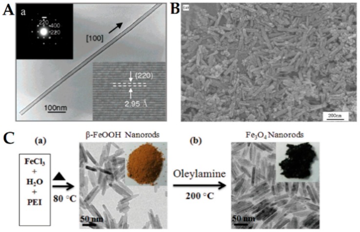
Figure 1.
(A) TEM image of a Fe3O4 nanotube. Upper inset: Selected area electron diffraction of the nanotube. Lower inset: HRTEM image taken on the tube wall. Reproduced from [85] with permission from American Chemical Society, 2020. (B) SEM image of magnetite nanorods described in [71]. Reproduced from [71] with permission from Elsevier, 2020. (C) Scheme of the two-step synthesis of magnetite nanorods. Adapted from [88] with permission from Royal Society of Chemistry, 2020.

