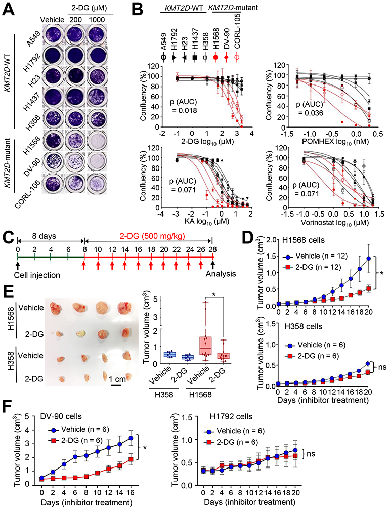
Figure 5. Inhibition of glycolysis suppresses tumorigenic growth of KMT2D-mutant LUAD cell lines.

(A and B) The effect of 2-DG on the confluency of human LUAD cell lines bearing WT KMT2D (A549, H1792, H23, H1437, and H358) and LUAD cell lines bearing KMT2D-truncating mutations (H1568, DV-90, and CORL-105) (A) and dose-response curves of several inhibitors on these LUAD cell lines (B). Cells were treated with various concentrations of the hexokinase inhibitor 2-deoxy-D-glucose (2-DG), the enolase inhibitor POMHEX, the GAPDH inhibitor koningic acid (KA), and the HDAC inhibitor SAHA/vorinostat. Wilcoxon rank sum test (n = 3) was used for statistical analysis. AUC, Area under the curve. (C–E) The effect of 2-DG on tumorigenic growth of H1568 cells bearing a KMT2D-truncating mutation and of H358 cells bearing WT KMT2D in a mouse subcutaneous xenograft model. The schedule of treatment of mice with 2-DG is shown (C). The sizes of xenograft tumors after treatment with 2-DG or vehicle control were measured (D). Tumors were dissected from the mice (E). In the boxplots, the bottom and the top rectangles indicate the first quartile (Q1) and third quartile (Q3), respectively. The horizontal lines in the middle signify the median (Q2), and the vertical lines that extend from the top and the bottom of the plot indicate the maximum and minimum values, respectively. (F) The effect of 2-DG on tumorigenic growth of DV-90 cells bearing a KMT2D-truncating mutation and of H1792 cells bearing WT KMT2D in a mouse subcutaneous xenograft model. Mice were treated with 2-DG (500 mg per kg body weight every other day) or vehicle. ns indicates non-significant. *, p < 0.05 (two-tailed Student’s t-test). See also Figures S5 and S6 and Table S3.
