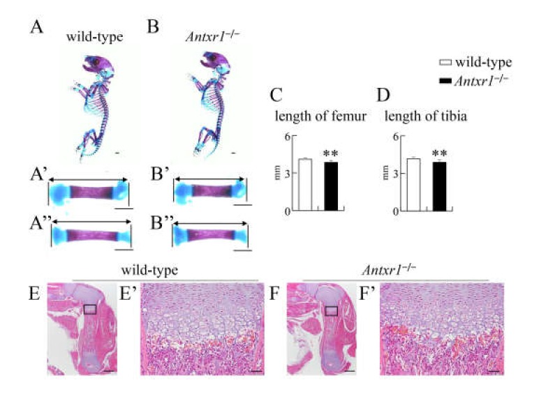
Figure 4.
Skeletal system and histological analysis of wild-type and Antxr1–/– newborns: (A,B) Lateral view of the whole skeletons (A,B) and magnified pictures of femurs (A’,B’) and tibiae (A’’,B’’) in wild-type (A) and Antxr1–/– (B) newborns. (C,D) The lengths of femurs (C) and tibiae (D): The lengths of both sides of femurs and tibiae were measured using skeletal preparations of 4 wild-type and 3 Antxr1–/– newborns, as shown in A’,B’,A’’ and B’’. Versus wild-type newborns, **p < 0.01. (E,F) H-E staining using femoral sections from wild-type (E) and Antxr1–/– (F) newborns. The boxed regions in E and F are magnified in E’ and F’, respectively. Scale bars: 1 mm (A,B), 500 µm (E,F), and 100 µm (E’,F’). The number of mice analyzed: H-E staining, wild-type: 2, Antxr1–/–: 2.

