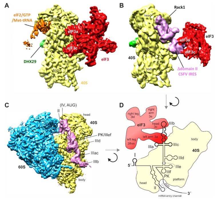
Figure 4.
Binding of the HCV IRES to the ribosome and to eIF3. (A) The small ribosomal 40S subunit (yellow) with bound eIF3 (red). eIF3 makes multiple contacts to the 40S subunit using several subunits, including eIF3a and eIF3c; (B) The IRES of classical swine fever virus (CSFV) (pink) without IRES domain II, binding to the 40S subunit (yellow) and to eIF3 (red). The IRES has displaced eIF3 completely from its binding to the 40S subunit (compare panel A) but keeps it connected to the 40S subunit only indirectly by contacts between IRES SL IIIabc and eIF3. Figures A and B were reprinted from [150] (Figure 2F) and slightly modified. Reprinted with permission from Elsevier (licence no. 4761840166773); (C) The HCV IRES (pink) binding to the 40S subunit (yellow) in the complete 80S ribosome (60S subunit in blue). The IRES SL IIIdef/PK is in close contact with the 40S subunit, the SLs II and IV are positioned in the mRNA entry channel, and the SL IIIb is pointing to the solvent side for binding eIF3. The IRES is not touching the 60S subunit [121,138]. Figure C was modified from the left panel of Figure 1C in [121]. Reproduced with permission from EMBO; (D) Schematic illustration of the HCV IRES binding to the 40S subunit (yellow) and to eIF3 (red) when eIF3 is largely displaced from the 40S subunit by the IRES, similar as in (B). The orientation of the 40S subunit is shown essentially top-down as compared with (A–C), viewed approximately from the solvent side.

