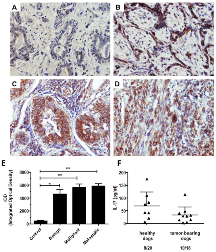
Figure 7.
The immunohistochemical expression of IL-17 in canine mammary tumors and the plasma levels of IL-17 in healthy and mammary tumor-bearing dogs. Representative light micrographs of canine mammary gland tissue (A), benign (B), malignant (C), and metastatic tumors (D) obtained with an Olympus BX60 microscope (at 200× total magnification). The IL-17 antigen is represented by the brown-colored precipitate in the cell cytoplasm and extracellular matrix. (E) The graph showing the IOD (integrated optical density) of IL-17 expression. The results are presented as the mean ± SEM. A one-way ANOVA and Tukey HSD post hoc test were applied. Significance levels are indicated as follows: * p < 0.05; ** p < 0.01. (F) The concentration of IL-17A (pg/mL) in the plasma of the client-owned healthy (8 out of 21) and malignant canine mammary tumor-bearing dogs (10 out of 18). Student’s t- test was applied, and no statistical significance was observed (p > 0.05).

