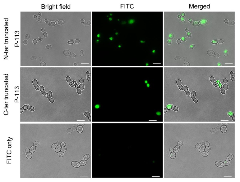
Figure 8.
Localizations of FITC truncated P-113 peptides in C. albicans. Fluorescence microscopy of C. albicans (107 CFU/mL) incubated at 28 °C for 30 min with 50 μM of FITC-truncated P-113 or FITC only. The left panels show a bright field, the middle panels show FITC images, and the right panels show merged images. The bar corresponds to 5 μm.

