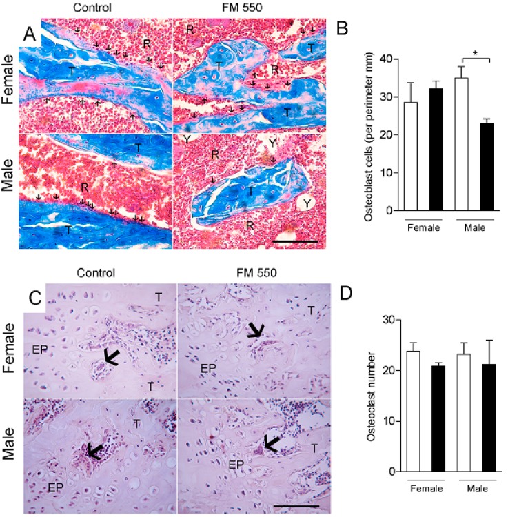
Figure 4.
Developmental exposure to FM 550 decreases osteoblast number in male but not female rats. Histomorphometric analysis of the femurs using Masson’s trichrome or tartrate resistant acid phosphatase (TRAP) staining. (A) Representative images of Masson’s trichrome staining in female (top) and male (bottom) rats exposed during development to FM 550. Black arrows indicate osteoblasts cells along the trabecular bone. T, trabecular bone. R, red bone marrow. Y, yellow bone marrow. Scale bar, 100 µm; (B) Mean ± SEM number of osteoblast. Two-way ANOVA followed by the Holm–Sidak post hoc test (n = 4 per group). * p < 0.05 different from control of the same sex; (C) Representative images of TRAP-positive osteoclasts in female (top) and male (bottom) rats from Control and exposed to FM 550 groups. Large arrows indicate osteoclasts. EP, epiphyseal disc. T, trabecular bone. Scale bar, 100 µm; (D) Mean ± SEM number of TRAP-positive osteoclasts. Two-way ANOVA followed by the Holm–Sidak post hoc test (n = 4 per group) determined no effect of sex (p = 0.95) or FM 550 (p = 0.29).

