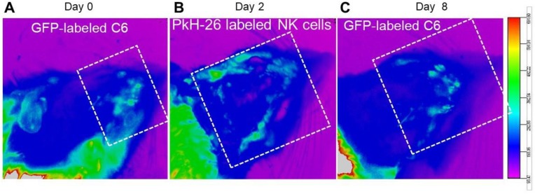
Figure 1.
Bio-distribution of NK cells in the rat model of induced GBM; live imaging. Bio-distribution of PKH-26-labeled NK cells 2 days after intravenous injection in our GFP-labeled GBM tumor induced model. (A) GFP-labeled GBM cells were found at day 0 (tumor establishment, white dotted frame). (B) Fluorescence imaging on day 2 shows the localization of PKH-labeled NK cells around GFP-labeled tumor cells (white dotted frame). (C) A reduction of fluorescence intensity of GFP-labeled GBM cells was evident 8 days post injection, indicating the effectiveness of the introduced NK cells (white dotted frame).

