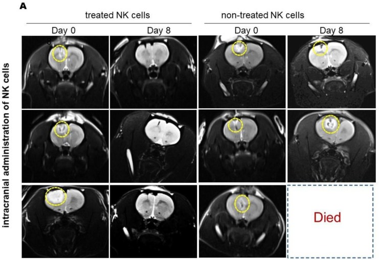
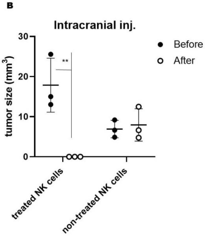
Figure 4.
MRI results of intracranial injection of NK cells in the rat model of induced GBM. (A) T2-weighted MR Images of GBM tumor-bearing rats show tumor shrinkage over time to nearly undetectable size at day 8, after injection with HSP70/IL-2-treated NK cells (panels on the left). No robust effects of injection with nontreated NK cells were observed (panels on the right) yellow dotted circles show tumor zones. (B) Graph depicting tumor volume shrinkage at day 8 in rats intracranially injected with treated NK cells versus nontreated NK cells (** p-value: 0.0319). Tumor size in missed (dead) cases was considered as unchanged.


