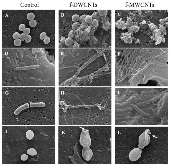
Figure 11.
SEM images of (A–C) Staphylococcus aureus, (D–F) Pseudomonas aeruginosa, (G–I) Klebsiella pneumoniae, and (J–L) Candida albican microbial cells. The images refer to the untreated control group and microbial cells exposed to 100 μg/mL f-DWCNTs and f-MWCNTs at 80,000 × magnification. The arrows indicate the CNTs’ web.

