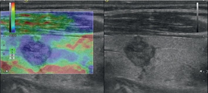
Figure 5.

Images in a 65-year-old woman who underwent routine check-up. A 10 mm left thyroid nodule with hypoechogenicity, solid, irregular margin, taller-than-wide, and no calcification was found by grayscale US and assessed as a malignant nodule. A score of 4 was assigned at elastography. The TI-RADS category was assigned as 5. This thyroid nodule was diagnosed as papillary thyroid carcinoma at surgery.
