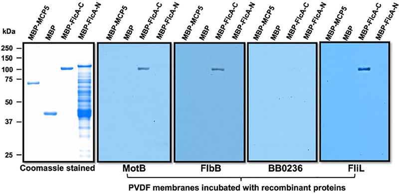
Figure 5. Specific interactions between FlcA and other flagellar proteins by affinity blotting.
Approximately 1 μg of MBP-tagged proteins shown on top of each panel were subjected to SDS-PAGE and then stained with Coomassie blue or transferred to a PVDF membrane. The membranes were incubated with 1xFLAG tagged MotB, FlbB, BB0236 or FliL, and then immunoblotted with anti-FLAG monoclonal antibodies.

