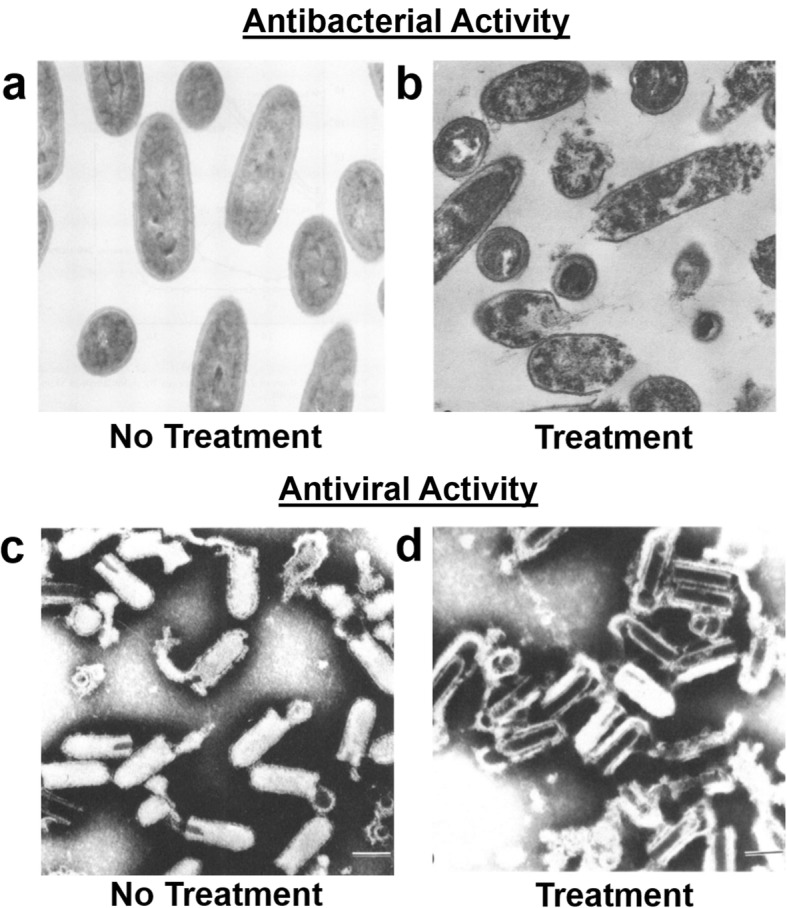
Fig. 1.

Membrane disruption of bacteria and enveloped viruses by free fatty acids and monoglycerides. a and b Transmission electron microscopy images of L. monocytogenes bacterial cells a without treatment and b after treatment with glycerol monolaurate. The magnification scale is × 44,080. Images are from Ref. [27] and reproduced with permission from the American Society of Microbiology. c and d Electron microscopy images of vesicular stomatitis virus particles c without treatment and d after treatment with long-chain linoleic acid (free fatty acid). Scale bars, 100 nm. Images are from Ref. [28] and reproduced with permission from the American Society of Microbiology
