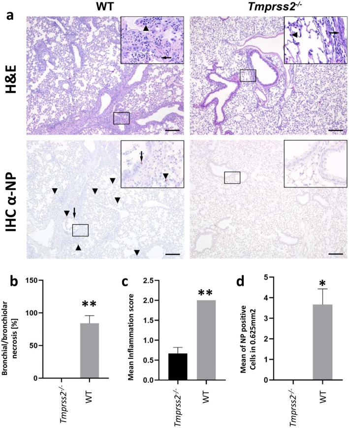
Fig. 3.
Strongly reduced lung tissue damage, immune cell infiltration and absence of virus replication in infected Tmprss2−/− mice. Female 8–12-week-old C57BL/6 J wild type (WT) and Tmprss2−/− knock-out (KO) mice were infected intranasally with 2 × 10⁴ focus forming units (ffu) PR8_HA(H2) (H2N1 virus (WT: n = 3; KO: n = 3). Lungs were prepared at 4 days post infection (dpi) and paraffin sections were stained with H&E (a top). Inserts: arrows point to damaged bronchiolar epithelium; arrowheads indicate mononuclear cells including lymphocytes and macrophages infiltrating lung interstitium and alveolar lumina adjacent to bronchioli. Bars, 200 μm. (a bottom) For detection of infected cells by immunohistochemistry, anti-NP (IHC α-NP) antibodies were used and sections were counterstained with Mayer’s hematoxylin. IAV-infected bronchiolar epithelial cells (arrow) and leukocytes (arrowhead) were found in WT mice, whereas no immunostaining was detectable in Tmprss2−/− mice (see inserts). b Necrotic bronchioli were determined as percentage of total cells. c Semi quantitative scoring results of cellular infiltration (0 = none, 1 = mild, 2 = moderate, 3 = severe). d IAV antigen positive cells were counted twice in 10 randomly selected high power fields (10 × 0.0625 mm2). Bars indicate mean values +/− SEM. One-sample t-tests were used for statistical analysis (* p < 0.05; ** p < 0.01). Please note that all values are identical for WT in the inflammation score (error bar is zero)

