
Figure 3.
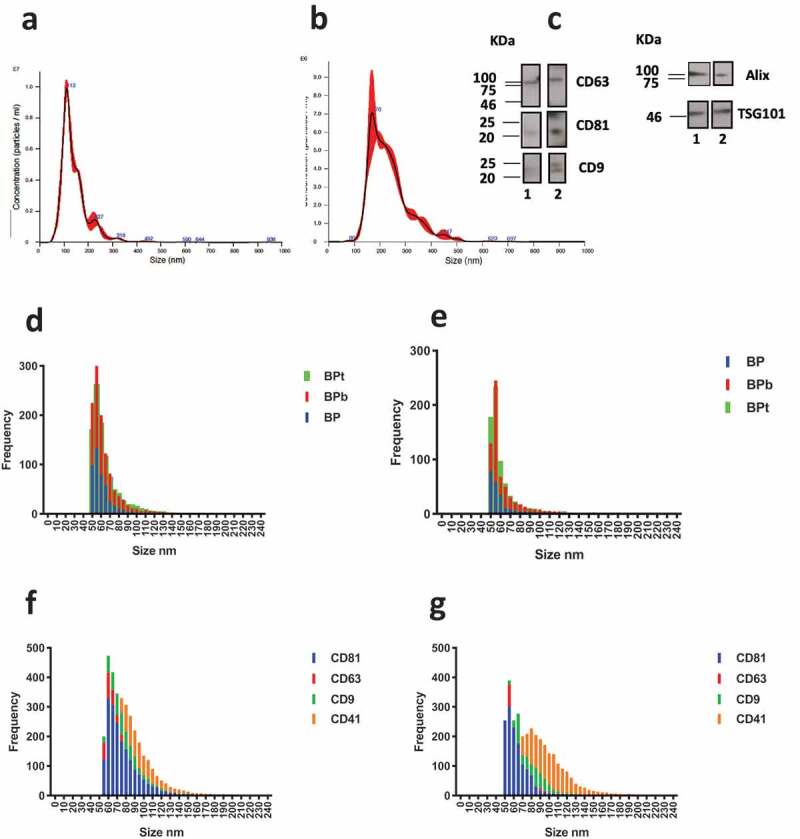
NTA analysis of (a) sEV-enriched sample obtained by centrifugation at 100.000 g for 2 h (mean particle diameter 143 ± 1 nm) and (b) MVs-enriched sample obtained at 10.000 g for 1 h (mean particle diameter 236 ± 5 nm). (c) Western blotting analysis for sEVs (lane 1) and MVs-enriched sample (lane 2). CD63, CD9 and CD81 markers are confirmed for both samples. Similarly, luminal proteins Alix and TSG101 are detected for both sEVs and MVs samples. (d) Observed size distribution on peptide chips for captured vesicles from the sEVs and (e) MVs-enriched sample obtained at 10.000 g. The size is reported as the number of counts detected in each 5 nm bin. (f) Size distribution on tetraspanins antibody chip for the sEVS sample and (g) MVs sample.