
Figure 4.
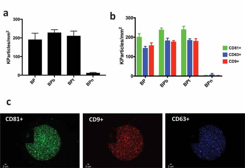
(a) EV density after incubation with HEK UC sample at the concentration of 1 × 10⁹ particles/mL label free detected on BP peptides; (b) correspondent EV density detected by fluorescence on BP peptides; (c) representative BP spot and fluorescence immune staining. Images were acquired on the three different fluorescence channels: green particles are vesicles captured by BP and positive for CD81; blue particles are vesicles captured by BP and positive for CD63 whereas red particles are vesicles captured by BP and positive for CD9.