
Figure 5.
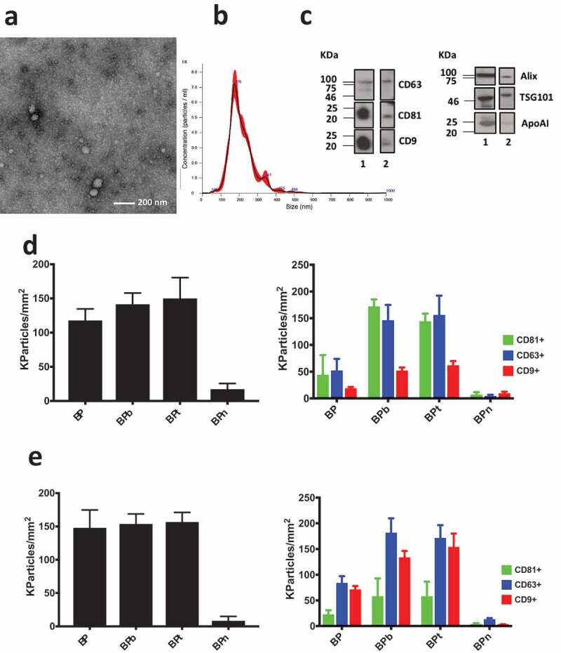
(a) TEM imaging of EVs obtained from human serum by UC followed by combined polymer precipitation and SEC showed results comparable with analogous analysis reported elsewhere [36]; (b) NTA analysis provided a mean particle size of 203 ± 3 nm and a concentration of 8.2 × 10¹¹ particles/mL; (c) the presence of transmembrane protein CD63, CD81 and CD9 and luminal proteins ALIX and TSG101 was assessed by Western blotting of serum (lane 1) and after combined isolation of EVs. Contamination by lipoproteins is assessed by WB of Apo AI in serum (lane 1) and in the purified EVs (lane 2). (d) Analysis of EV isolated by ultracentrifugation, polymer precipitation and SEC from human serum incubated on peptide microarrays at 1 × 10⁹ particles/mL concentration. Density of particles captured by BP peptides (left panel) is confirmed by fluorescence staining using CD81/CD/63/CD9 fluorescent antibodies (right panel). (e) Analysis performed on unpurified human serum diluted 1:8. Density of particles captured by BP peptides is detected label free (left panel) and by fluorescence staining using CD81/CD/63/CD9 fluorescent antibodies (right panel). Observed size distribution of captured EV is reported in the supplementary information (Figure S3).