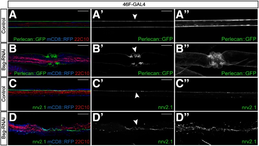
Figure 4.
Perineurial glial constriction causes constriction of both the overlying ECM and underlying septate junctions. A, B, Peripheral nerves of third instar larvae expressing endogenously tagged ECM component Perlecan::GFP (green) and 46F-GAL4 driving expression of mCD8::RFP (blue) with axons labeled using anti-Futsch/22C10 (red). Bsg knockdown in the perineurial glia results in compression of the ECM in regions of glial constriction (arrowhead). Projections of 3D-reconstructed nerves (B′′) show convoluted infolding of the ECM in 46F>Bsg-RNAi animals compared with the smooth sheath seen in control nerves (A′′). C, D, Peripheral nerves from 46F>mCD8::RFP larvae labeled using an antibody against core septate junction component Nervana 2.1 (nrv2.1, green). Septate junctions run laterally along the length of the nerve between the opposing membrane of the subperineurial glia in control animals (C′, arrowhead). In regions of glial compression, the septate junction strands appear convoluted (D′, arrowhead), but 3D reconstruction shows that the strands remain continuous (D′′). Scale bars, 15 μm.

