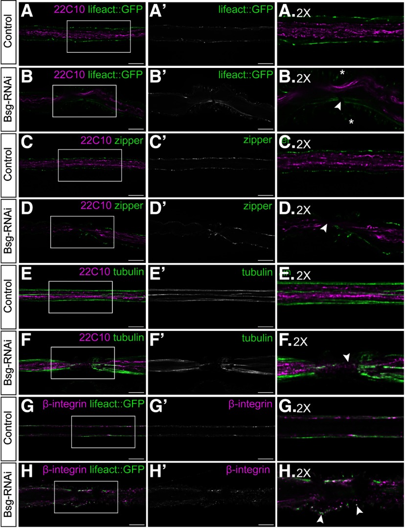
Figure 6.
Glial compression is associated with discontinuities in cytoskeleton components. A, B, Peripheral nerves of third instar larvae expressing filamentous actin marker lifeact::GFP (green) driven by 46F-GAL4, with axons labeled using anti-Futsch/22C10 (magenta). Actin filaments show discontinuities in the regions of glial compression (B, arrowhead), and lifeact::GFP-positive puncta are accumulated at the tips of the compressions (asterisks). C, D, Nerves labeled with anti-Futsch (magenta) and an antibody against nonmuscle myosin II (zipper, green) to show distribution of myosin motors. Regions of glial compression were associated with gaps in labeling of myosin (D, arrowhead). E, F, Nerves labeled with anti-Futsch (magenta) and anti-tubulin to show morphology of microtubules (green). In regions of glial compression, microtubules appear fragmented (F, arrowhead). G, H, Nerves expressing lifeact::GFP (green) and labeled with an antibody against the β-subunit of the integrin heterodimer (magenta). Integrin is localized to the focal adhesion complexes, which are distributed uniformly along the length of the glial sheath. In regions of glial compression, β-integrin is found within the compressed glial cells and at the tips of some the compressions alongside broken actin filaments (H, arrowheads). Side panels represent 2× digital magnifications of the indicated regions in the corresponding panels. Scale bars, 15 μm.

