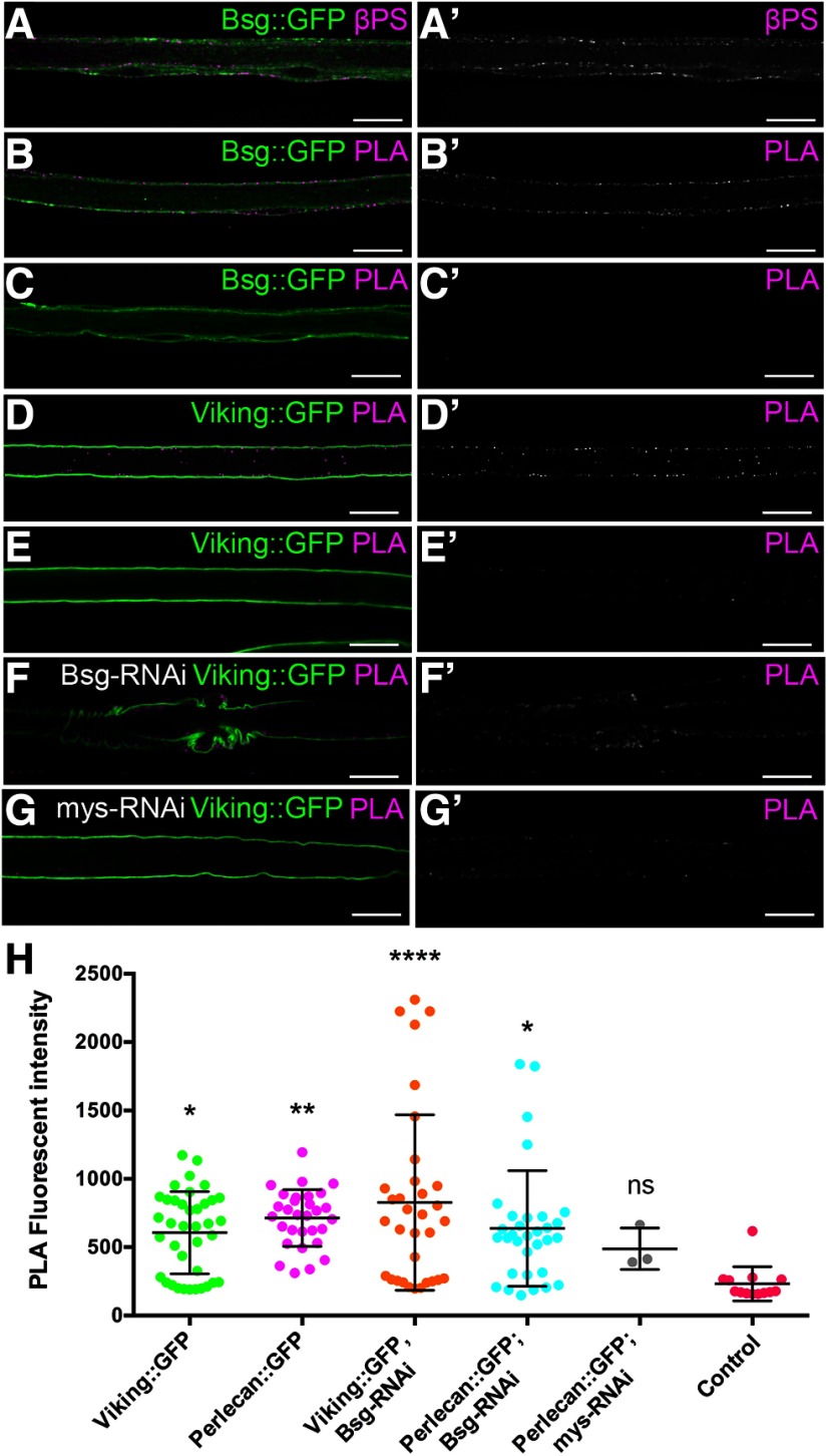
Figure 7.
Integrin is in close proximity to the extracellular domain of Bsg and the ECM in the perineurial glia. A, Peripheral nerve of Bsg::GFP larvae with integrin β-subunit labeled using anti-βPS (magenta). Bsg and integrin are coexpressed in the perineurial glia (arrowheads). B-J, PLA (magenta) with GFP tagged proteins (green) and the βPS integrin subunit. B, PLA using anti-GFP and anti-βPS antibodies shows that Bsg::GFP and integrin are found in close proximity (<40 nm) in the perineurial glia. C, Negative control of PLA experiment where anti-GFP antibody was absent shows no PLA. D, Positive control of PLA experiments showing ECM component collagen-IV (Viking::GFP) is found in close proximity to integrin. E, Negative control where the anti-GFP antibody was absent shows no PLA. F, Positive PLA between collagen-IV (Viking::GFP) and integrin occurs in knockdown of Bsg in the perineurial glia (46F>Bsg-RNAi). G, Control where β-integrin was knocked down in the perineurial glia (46F>mys-RNAi) shows reduced PLA with Viking::GFP. Scale bars, 15 μm. H, Quantification of the PLA reaction fluorescence intensity between the βPS integrin subunit and ECM components Viking and Perlecan tagged with GFP. Fluorescence intensity was significantly higher for all reactions compared with control (Viking::GFP minus the anti-βPS antibody) with the exception of when β-integrin (mys-RNAi) was knocked down. From left to right: Viking::GFP+βPS, *p = 0.0208; Perlecan::GFP+βPS, **p = 0.0023; Viking::GFP+βPS, Bsg-RNAi, ****p < 0.0001; Perlecan::GFP+βPS, Bsg-RNAi, *p = 0.0130; Perlecan::GFP+βPS, mys-RNAi, ns p = 0.7835. *p < 0.05, **p < 0.01.

