Figure 2.
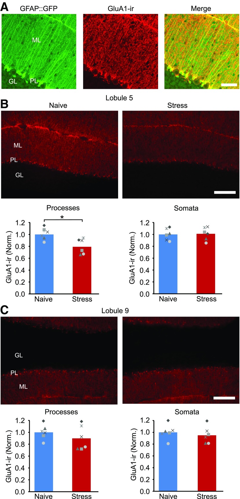
Stress reduces GluA1 expression in Bergmann glial cell processes in cerebellar lobule 5. A, Confocal images of cerebellar cortex stained for the GluA1 AMPA receptor subunit in GFAP::GFP mice shows that GluA1 is highly expressed in Bergmann glial cells. B, Top, Epifluorescence GluA1-IR images of lobule 5 in a naive control mice and after stress. Bottom, The mean intensity of GluA1-IR in Bergmann cell processes (in the molecular layer of the cerebellar cortex) and the somata of Bergmann cells (located in the Purkinje cell layer) in lobule 5. Stress reduced the level of GluA1-IR in the molecular layer (naive, N = 6; stress, N = 6). C, Top, GluA1-IR images of lobule 9. Bottom, Mean GluA1-IR in Bergmann processes and somata in cerebellar lobule 9. ML, Molecular layer; GL, granule cell layer; PL, Purkinje cell layer; ns, not significant. Scale bars: A, 50 µm; B, 100 µm. *p < 0.02 (unpaired t test).

