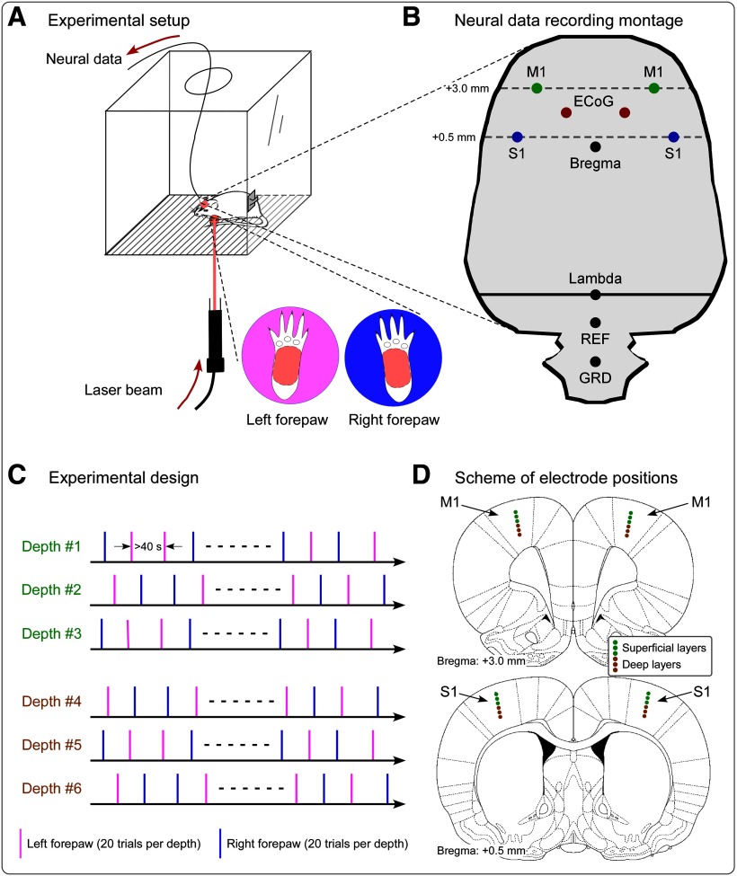
Figure 1.
Experimental design and recording setup. A, During the recording sessions, rats were free to move within a plastic chamber (30 × 30 × 30 cm3). When the animal was spontaneously still, laser stimuli were delivered on the plantar surface of either the left or the right forepaw through gaps on the floor of the chamber. C, In each recording session, the neural activity was measured at one of six cortical depths. In each session, we delivered 20 laser pulses to the right forepaw and 20 laser pulses to the left forepaw, in pseudorandom order. The interval between two consecutive stimuli was never <40 s. B, D, Scheme showing the position of electrodes for the simultaneous intracortical and epidural recording. B, Positioning of the four microelectrodes for the recording of LFPs and single-unit activity in the S1 and M1 contralateral and ipsilateral to the stimulation site, as well as of epidural electrodes. Intracortical microelectrodes were placed according to stereotaxic coordinates in the following positions [expressed in respect to the bregma (in mm); positive x-axis and y-axis values indicate right and anterior locations, respectively]: left S1: x = −4, y = 0.5; right S1: x = 4, y = 0.5; left M1: x = −3, y = 3; right M1: x = 3, y = 3. Epidural electrodes were placed in the following positions: left ECoG: x = −1.5, y = 1.75; right ECoG: x = 1.5, y = 1.75. Reference (REF) and ground (GRD) electrodes were placed 2 and 4 mm caudally to the lambda, on the midline. D, Intracortical neural data were recorded at six different depths. Electrode positions measuring data from superficial and deep cortical layers are marked in light green and brown, respectively.

