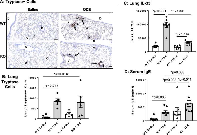
Fig. 5.
Tryptase+ lung mast cells, serum IgE and lung IL-33 levels are increased following repetitive ODE exposure and variably modulated by MyD88. WT and MyD88 KO mice were treated i.n. daily for 3 weeks with saline or ODE whereupon lung sections (5 μm thick) were stained for tryptase with representative images for each treatment group shown a. Key: b, bronchiolar airway; a, alveolar parenchyma; v = blood vessel; asterisk, cellular aggregates; and arrow denotes positive tryptase stained cells in cellular aggregates (scale bar is 100 μm). b, Scatter plot depicts means with SEM of tryptase positive cells per entire lung section as quantified by Definiens software with N = 5–6 mice/group. c, Serum IgE levels quantified by ELISA with N = 8–9 mice group. d, ODE-induced lung homogenate IL-33 levels were reduced in MyD88 KO mice with N = 6–7 mice/group

