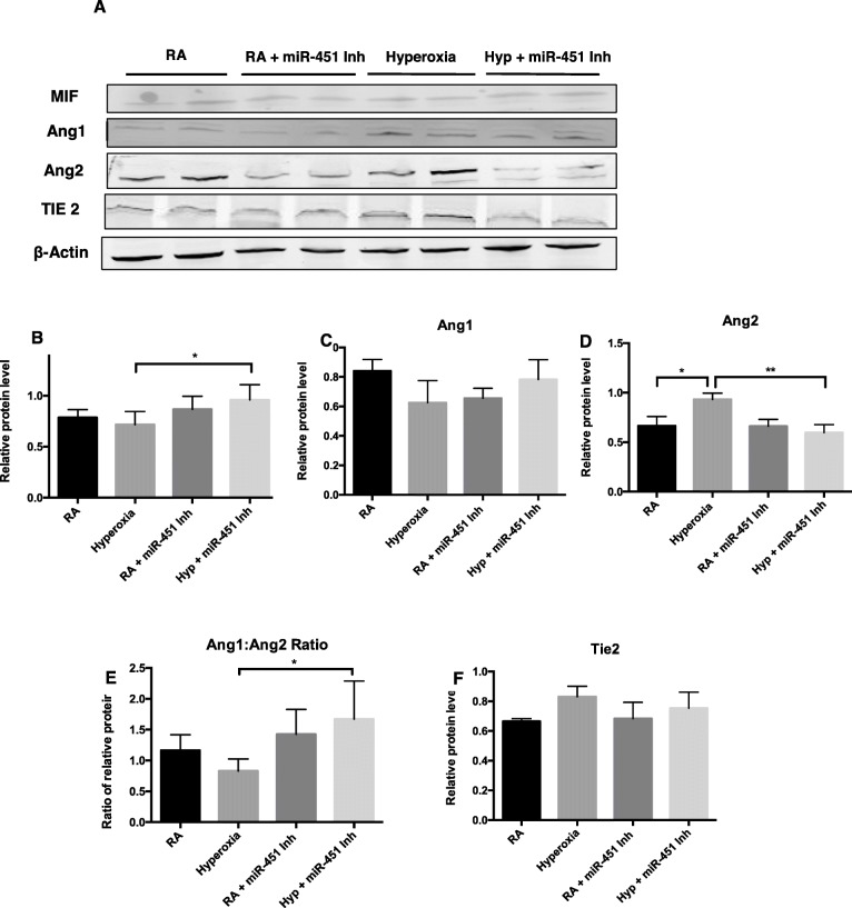
Fig. 2.
Inhibition of miR-451 is associated with increased expression of MIF in MLECs exposed to hyperoxia. a Representative image of Western blot analysis showing expression of macrophage migration inhibitory factor (MIF), Angiopoietin 1 (Ang 1), Ang 2, and their receptor, Tie 2 and β-actin in MLECs exposed to hyperoxia and transfected with a miR-451 inhibitor. b MIF expression quantified by densitometry with normalization to β-actin. c and d Expression of the vascular growth factors Ang1, Ang2 quantified by densitometry with normalization to β-actin. e Ratio of the densitometric values of Ang1 and Ang2. f Expression of the Ang receptor Tie2 quantified by densitometry with normalization to β-actin. N = 3–4, in each group. RA: room air, Hyp: hyperoxia 95% O2 for 16 h, miR-451 Inh: miR-451 inhibitor. * p < 0.05 **P < 0.01

