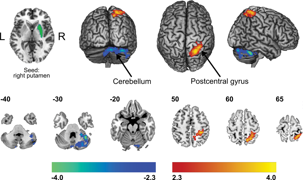
Figure 3. Group differences in resting state functional connectivity of the putamen.
Compared to matched controls, CRPS patients demonstrated greater functional connectivity strength (warm colors) amongst the right (ipsilateral to the affected limb) putamen and sensorimotor and superior parietal cortices, while decreased connectivity (cold colors) was quantified with the crus I region of the cerebellum. Statistical maps were thresholded at p-value < 0.05, corrected for multiple comparisons. Color bars show z-values.

