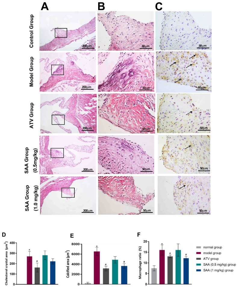
Figure 4.
SAA ameliorated histopathological changes in the aortic tissues of T2DM ZDF rats with AS. The ZL and ZDF rats were sacrificed, and the aortic tissues were collected for H&E staining (A line, 200×. B line, 400×) as well as IHC staining (C line, 400×). Representative activated macrophages in the aorta were stained with anti-CD68 and are indicated by black arrows. Histopathological quantitative analysis was performed by experienced pathologists. The data of cholesterol crystal area (D), calcified area (E) and macrophage ratio (F) are shown. Data are expressed as mean ± SEM. * p < 0.05 vs. normal group (Zucker lean control rats). # p < 0.05 vs. model group (Zucker diabetic fatty rats with HFD and VD3 injections).

