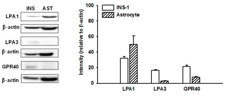
Figure 2.
Expression of LPA1, LPA3, and GPR40 in INS-1 cells and mouse astrocytes. Cell lysates were subjected to immunoblotting using antibodies against LPA1, LPA3, and GPR40. The expression of each receptor relative to β-actin was plotted. Data represent means ± SD (n = 4). INS, INS-1 cells; AST, mouse astrocytes. LPA: lysophosphatidic acid.

