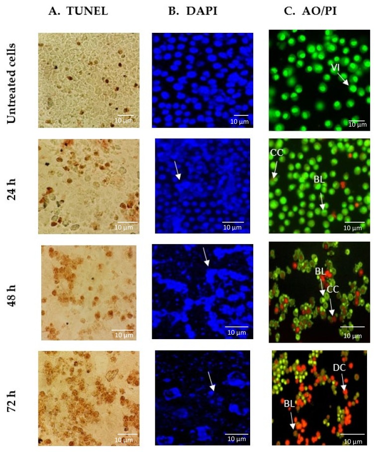
Figure 3.
Images of GNST-ITC-induced cell death in MCF-7 cells following incubation for 24, 48, and 72 h. (A) Micrograph of TUNEL assay under bright field microscopy with darkened stains indicating DNA fragmentation within the cells. (B) Alteration in nuclear morphology of GNST-ITC-treated MCF-7 cells evaluated using DAPI staining with arrows indicating chromatin condensation in the cell nucleus. (C) Fluorescence image of MCF-7 cells treated with AO/PI double staining with arrows indicating viable cells (VI), chromatin condensation (CC), membrane blebbing (BL), apoptotic bodies (AB), and dead cells (DC). Results are representative of three independent experiments. Magnification ×400.

