Figure 1.
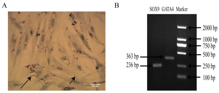
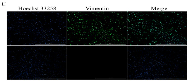
Identification of Sertoli cells. (A) The short arrow indicates bipolar bodies and the long arrow indicates lipid droplets, at ×40 magnification. (B) Expression of SOX9 and GATA4 mRNA. (C) The nucleus was counterstained with Hoechst 33258 and green fluorescence indicates vimentin positive (×4 magnification). N = 3 for both.


