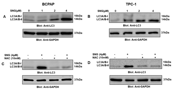
Figure 6.
Induction of autophagy in PTC cells treated with SNG. BCPAP (A) and TPC-1 (B) cells were treated with increasing doses of SNG for 4 h, as indicated. After cell lysis, equal amounts of proteins were separated by SDS–PAGE, transferred to a PVDF membrane, and immunoblotted with antibodies against LC3 and GAPDH. NAC pretreatment of PTC cells prevented SNG-induced LC3 expression. BCPAP (C) and TPC-1 (D) cells were pretreated with 10 mM NAC and subsequently treated with 4 µM SNG for 4 h. Cells were lysed, and 50 μg of proteins separated on SDS-PAGE, transferred to a PVDF membrane, and immunoblotted with antibodies against LC3 and GAPDH.

