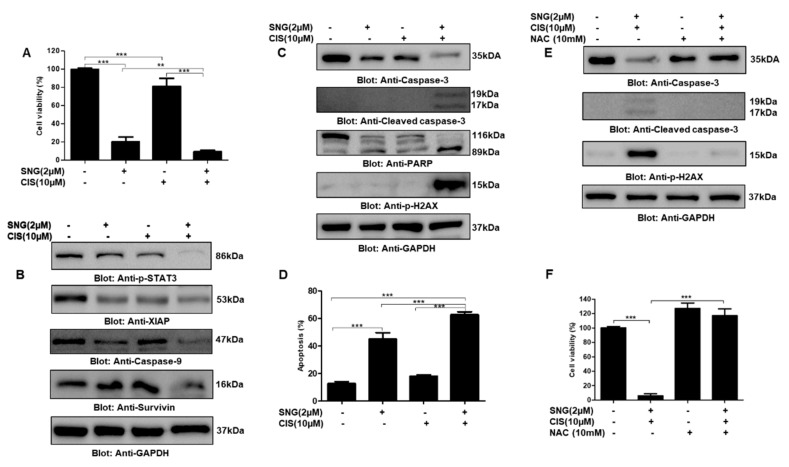
Figure 7.
Sanguinarine sensitized PTC cells to anticancer drug cisplatin. (A) TPC-1 cells were treated with 2 µM SNG and 10 µM of cisplatin alone and in combination for 24h. CCK-8 was used to determine cell viability, as described in materials and methods. Values are expressed as the mean +/− SD (standard deviation) of at least six replicates with p value ** p < 0.01, *** p < 0.001 (n = 6). (B,C) The combination of SNG and cisplatin inhibits STAT3 phosphorylation and induces apoptosis: TPC-1 cells were treated with 2 µM SNG and 10 µM of cisplatin alone and in combination for 24 h, cells were lysed and separated by SDS-PAGE, transferred to a PVDF membrane, and immunoblotted with antibodies against p-STAT3, XIAP, caspase-9, survivin, caspase-3, cleaved caspase-3, PARP, p-H2AX and GAPDH. (D) SNG and cisplatin augmented apoptosis in PTC cells: TPC-1 cells were treated with 2 µM SNG and 10 µM, alone and in combination, for 24 h and processed using annexin V and dead cell kit, according to the manufacturer’s instructions and analyzed by Muse® cell analyzer, as described in materials and methods. The graph displays the mean ± SD (standard deviation) p < 0.001 (n = 3). ROS plays an important role in SNG-mediated sensitization of PTC cells to cisplatin: (E) TPC-1 cells were treated with 10 mM NAC, 2 µM SNG and 10 µM of cisplatin, alone and in combination as indicated, for 24 h. Cells were lysed, and proteins were separated on SDS-PAGE, transferred to a PVDF membrane, and immunoblotted with antibodies against caspase-3, cleaved caspase-3, p-H2AX and GAPDH. (F) TPC-1 cells were treated with 10 mM NAC, 2 µM SNG and 10 µM of cisplatin, alone and in combination as indicated, for 24 h. CCK-8 was used to determine cell viability, as described in materials and methods. Values are expressed as the mean +/− SD (standard deviation) of at least six replicates (n = 6). *** p < 0.001.

