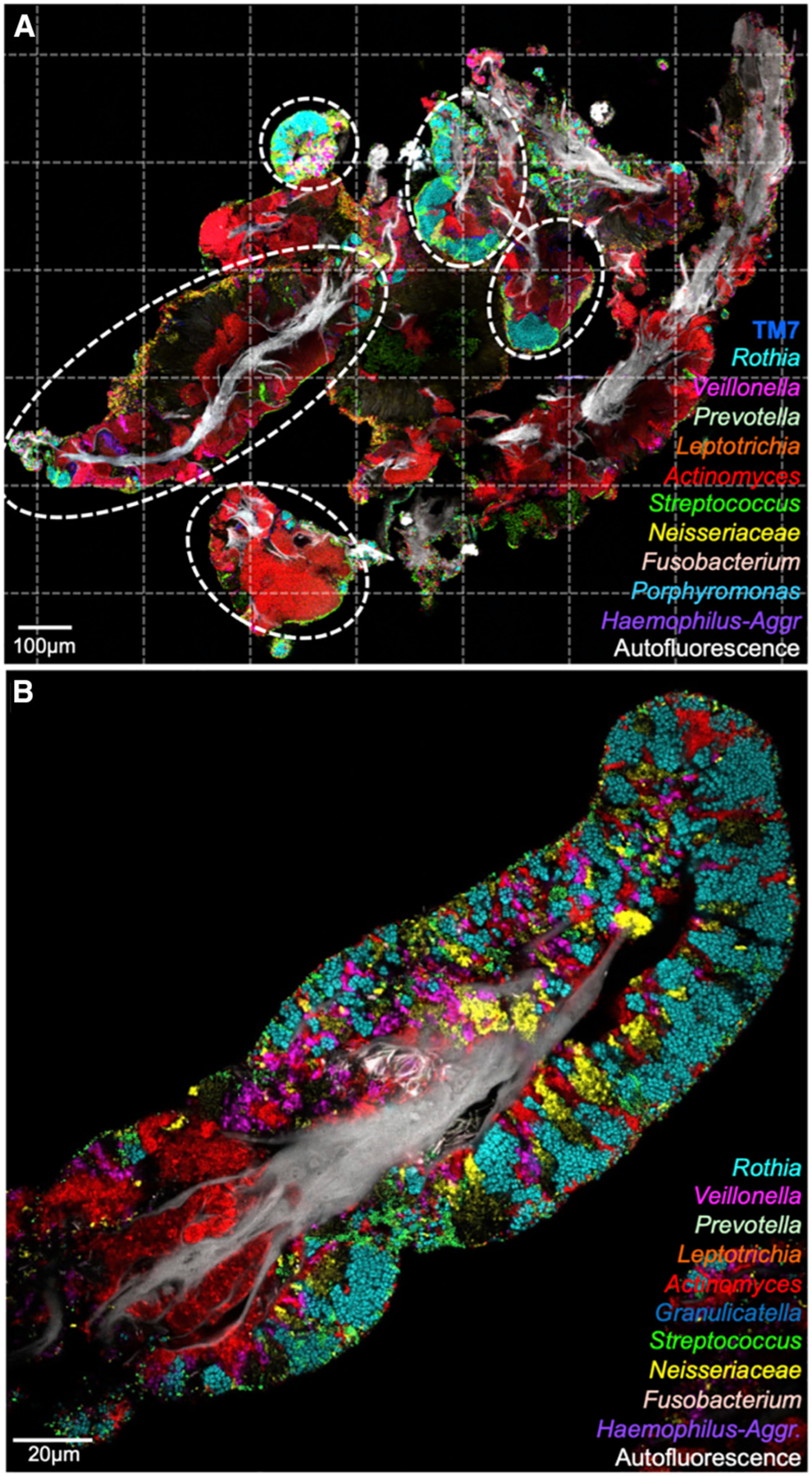
Figure 3. Tongue Biofilm Consortia Are Complex and Structurally Organized.

(A) Tile-scanned view (63 tiles; dotted lines) of material sampled from the TD. Host epithelial cells identified by autofluorescence are shown in white; colors indicate genera of bacteria. Bacterial consortia (circled) range in size from fifty to hundreds of microns.
(B) The spatial organization of an isolated consortium. Clusters of Rothia, Veillonella, Actinomyces, Neisseria, and Streptococcus cells comprise a large fraction of the consortium’s bacterial biomass. See also Figure S3 and Tables S2, S3, and S4.
