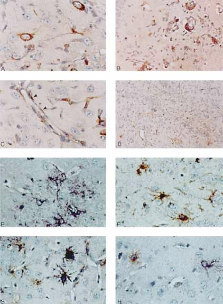
Fig. 1.
A-D Immunohistochemical analysis of MHV-A59-infected β2M(-/-) mouse brains. A Neuronal infection is exhibited by positive immunostaining for MHV antigen in the cytoplasm of parahippocampal neurons. Note the morphological stigmata of neurons: the large cell size, the characteristic oval nucleus with the prominent nucleolus, and the thick axonal and dendritic processes. × 400. B Viral antigen detected in perivascular lymphocytes (arrowheads). × 200. C Viral antigen detected in endothelial cells delineating cerebral small venules and capillaries (arrowheads). × 400. D Area of prominent rod-shaped microglial cell proliferation including many that stain positively for viral antigen. × 100. E-H Combined in situ hybridization for viral genome and GFAP immunohistochemical staining for astrocytes. E GFAP-negative neuronal cells expressing viral genome (purple). Small purple dots represent positive viral genome in cross-sectioned cell processes. × 400. F Uninfected reactive astrocytes which are negative for viral genome by in situ hybridization expressing abundant GFAP by immunohistochemistry. × 400. G Infected astrocytes (arrows) exhibiting both GFAP (by immunohistochemistry with GFAP antibodies) and viral genome (in situ hybridization with MHV-specific probes). × 400. H A neuron expressing viral genome by in situ hybridization (purple) next to a GFAP-positive astrocyte (brown). × 400.

