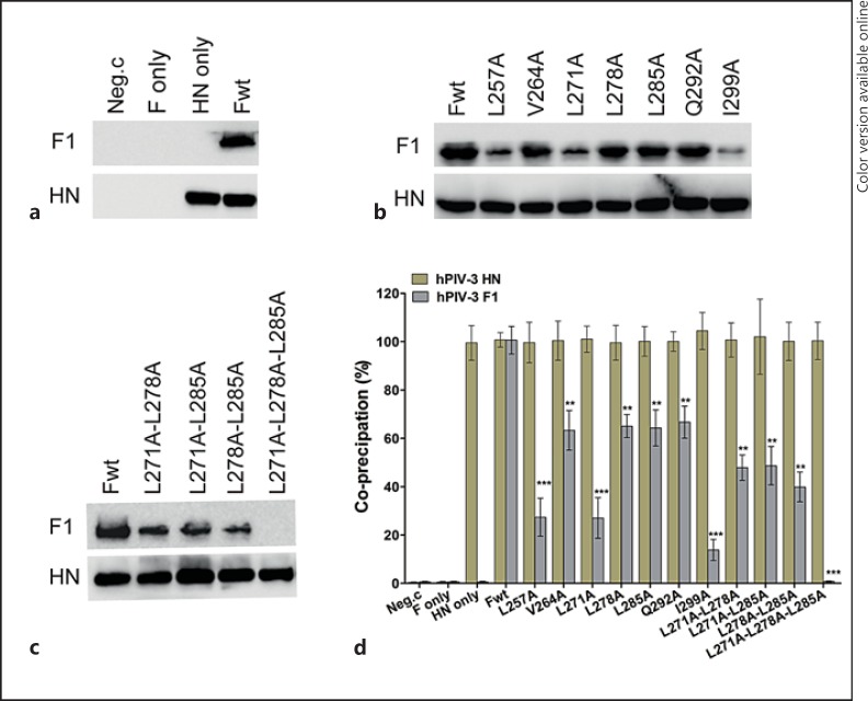
Fig. 7.
Coimmunoprecipitation of hPIV-3 wt or mutated F proteins with hPIV-3 attachment proteins. Surface proteins of cells transfected with the desired plasmids were biotinylated, and then the cells were lysed. Dynabeads-HN Ab-HN Ag-F Ag immune complexes were formed with the Dynabeads-cross-linked anti-HN antibody; after being washed sufficiently, they were resuspended in PBS and then incubated with streptavidin beads as described in Materials and Methods to precipitate the cell surface complexes. The coimmunoprecipitated F proteins in the complexes were detected by Western blot analysis with the anti-F monoclonal antibody (top), while the HN proteins were detected using a mixture of anti-HN monoclonal antibodies (bottom). All proteins were electrophoresed in the presence of a reducing agent. a Critical controls in this coimmunoprecipitation assay: Neg.c = Empty vector; HN only and F only = these plasmids alone; Fwt = wt F protein and HN protein. b, c The results of a coimmunoprecipitation assay for each of the proteins carrying alanine substitutions in the leucine zipper-like motif. wt F protein or the mutants indicated were cotransfected with hPIV-3 HN and are indicated above the gels. d Quantification analysis for b and c using the densitometry method from 3 independent experiments. Data are shown as means ± SD. ** p < 0.01, *** p < 0.001.

