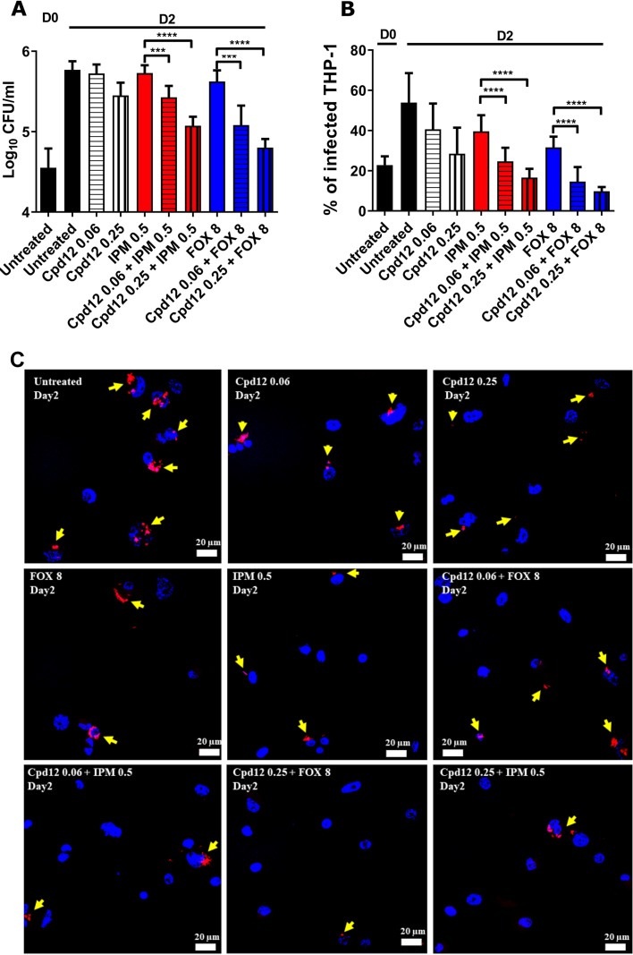
FIG 4.
Impact of Cpd12 alone or in combination on intracellular-residing M. abscessus. THP-1 macrophages were infected with M. abscessus S expressing TdTomato (multiplicity of infection [MOI] of 2:1) and treated with the indicated compounds (μg/ml). (A) CFU were determined at day 0 and day 2 postinfection. Data represents the mean ± SD of three independent experiments completed in triplicate. For statistical analysis, a nonparametric Mann-Whitney t test was performed. ***, P ≤ 0.001; ****, P < 0.0001. (B) Percentage of infected THP-1 macrophages at day 0 and day 2 postinfection. Data shown are mean values ± SD for one representative experiment completed in triplicate. One-way analysis of variance (ANOVA) Kruskal-Wallis was used as a statistical test. ****, P < 0.0001. (C) Immunofluorescent fields were taken at day 2 postinfection at magnification 40× (using confocal microscopy) showing the nuclei of macrophages (DAPI in blue) infected with red-fluorescent M. abscessus in the absence or in the presence of the drugs used alone or in combination. Yellow arrows emphasize red-fluorescent M. abscessus (tdTomato) within macrophages. Only intracellular bacteria that were individually observed under the microscope were recorded.

