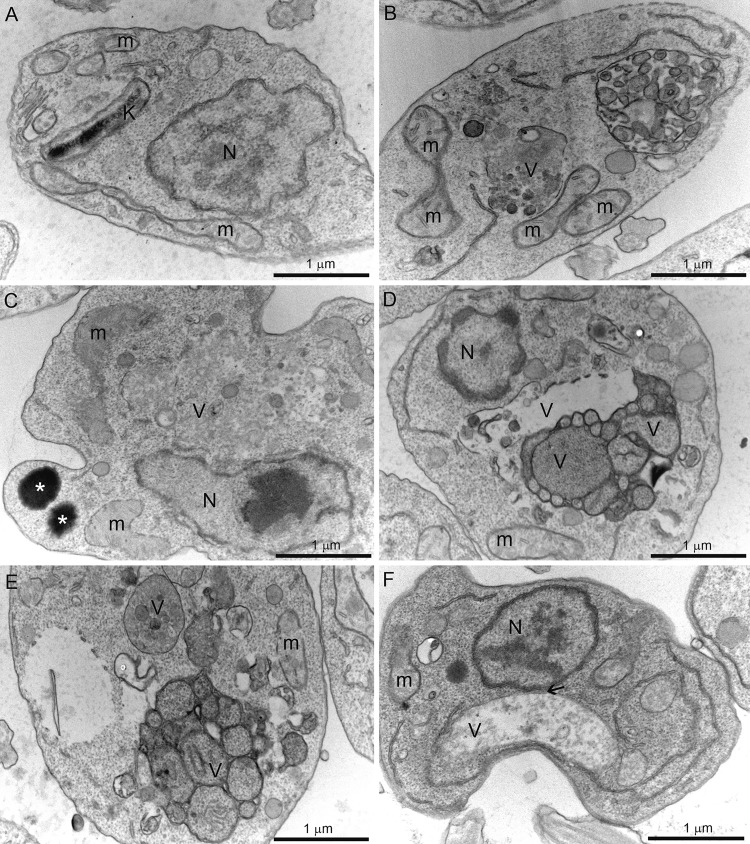
FIG 7.
Transmission electron microscopy of Leishmania amazonensis promastigotes control (A) and treated with 0.5 μM itraconazole (ITZ) (B, C), 0.5 μM compound 1 (D, E), or 0.5 μM compound 2 (F) after 72 of treatment. Several alterations were observed, such as intense disorganization and swelling of the mitochondrion (B, C, E), the presence of lipid bodies (C), and the appearance of vacuoles similar to autophagosomes (B–F) close to organelles such as the nucleus (F, arrow). V, vacuole; K, kinetoplast; M, mitochondrion; N, nucleus.

