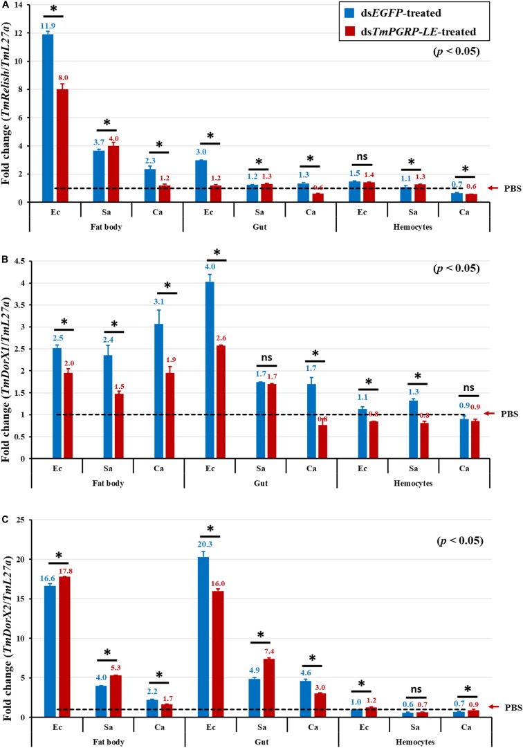
FIGURE 7.
Transcriptional activation of different NF-κB genes in the fat body, hemocytes, and gut of TmPGRP-LE dsRNA-injected T. molitor larvae after inoculation with E. coli (Ec), S. aureus (Sa), and C. albicans (Ca) (n = 20 per group). mRNA quantities of TmRelish (A), TmDorX1 (B), and TmDorX2 (C) in TmPGRP-LE knockdown larvae were measured relative to those for L27a at 24 h post-challenge by qRT-PCR. EGFP RNAi was used as a negative control. Bars represent mean ± SE of three independent experiments and the numbers above the bars indicate the transcription levels of NF-κB genes. Significant differences between dsEGFP- and dsTmPGRP-LE-treated groups are presented by asterisks (*) (p < 0.05) and ns, not significant.

