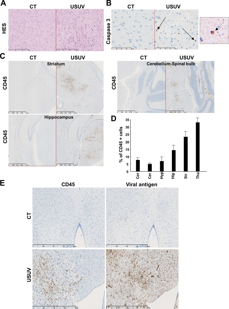
Fig 3. USUV induces apoptosis and cellular infiltration in the mouse brain.
(A) Transverse sections of the brain (thalamus) of USUV-infected and control (mock, PBS injected) mice at 6 dpi stained with haematoxylin/eosin/saffron. We observe cellular infiltration and cell shrinkage. CT = control mice. (B) Some cells present nuclear condensation and caspase 3 staining after immunohistochemistry, suggesting apoptotic process in USUV-infected mice. Right panel: zoom on apoptotic cell. Arrows indicate apoptotic cells. (C) Immunohistochemical CD45 staining (associated with luxol blue) showing massive inflammatory infiltrates in multiple areas of the infected brain. (D) Quantification of CD45 positive cells in different brain area. Cor: Cortex, Cer: Cerebellum, Hyp: Hypothalamus, Hip: Hippocampus, Str: Striatum, Tha: Thalamus. (E) Immunohistochemical staining of viral antigen and CD45. The majority of infected cells are not inflammatory cells.

