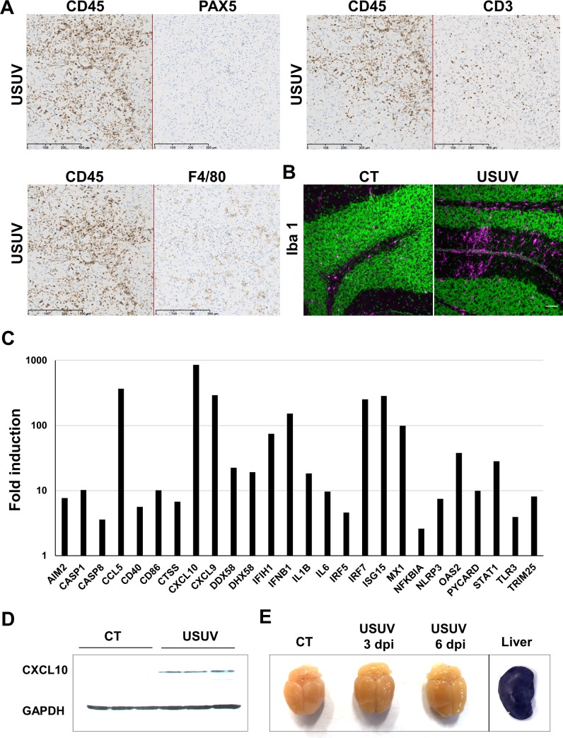
Fig 4. USUV induces massive brain inflammation.
Histological sections of neonatal brain mice infected by USUV (6 dpi). (A) Immunohistochemical staining of B-cells (PAX 5), T-cells (CD3) and macrophages (F4/F80) showing T-cells and macrophages infiltration. (B) Immunofluorescence staining (Iba1 in pink, nucleus in green) shows microglia recruitment/activation in USUV-infected brain contrary to control mice (CT). (C) RT-qPCR analysis using a specific inflammatory cytokines and receptors PCR array of mRNA collected at 6 dpi from brain of control and infected mice. Fold regulation of statistically significant genes normalized to CT are indicated for 3 mice of each group. p <0.05 (p value in S3A Fig). Scale bar = 50μm. (D) CXCL10 protein level in control and infected mice. (E) Representative picture of dissected brain from EB-injected mock- and USUV- infected mice at 3 and 6 dpi showing no blue labelling. Liver is used as positive control of EB staining.

