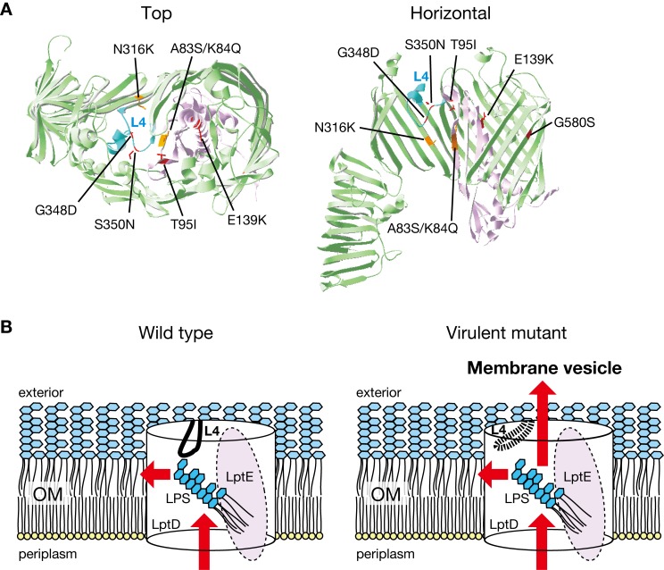
Fig 8. Location of the LptD and LptE mutations in the LptD-LptE complex structure.
(A) The structural location of the LptD and LptE mutations that increase E. coli virulence is presented. The LptD-LptE complex structure is from S. flexneri (4rhb). The left image is from the exterior side, and the right image is from the horizontal side. In the right image, LptD 635–784 was removed for clarity. Loop 4 of LptD is shown in cyan, the other parts of LptD are shown in moss green, and LptE is shown in purple. The mutations obtained in the experimental evolution are in red, and the mutations present in clinical isolates are in orange. (B) Model for structural alteration and LPS translocation in the LptD and LptE mutants. In the wild-type strain, loop 4 plugs the LPS tunnel and assists in the translocation of LPS into the outer membrane (left). In the LptD and LptE mutants, the structure of loop 4 is changed, thereby attenuating its plug function and allowing LPS to be secreted into the extracellular milieu (right).

