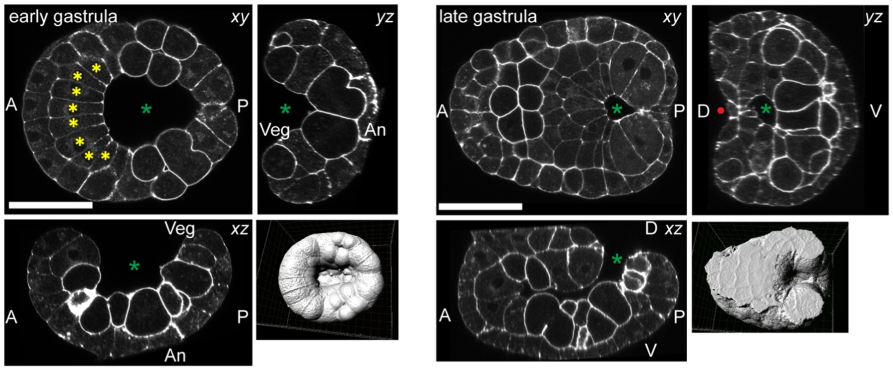
Figure 2. Ciona gastrulation.

Confocal images of gastrulating Ciona robusta embryos at the stages indicated. The panels on the left are derived from confocal stacks of a phalloidin-stained embryo early in gastrulation, with orthogonal xy, xz and yz sections shown. The surface-rendered view of the stack is from the vegetal side. The green asterisk marks the open blastopore/archenteron. Yellow asterisks mark notochord precursor cells. The panels on the right show an older embryo near the end of gastrulation/beginning of neurulation. The red dot indicates the neural groove just starting to form on the posterior dorsal side. Scale bars=50μm.
