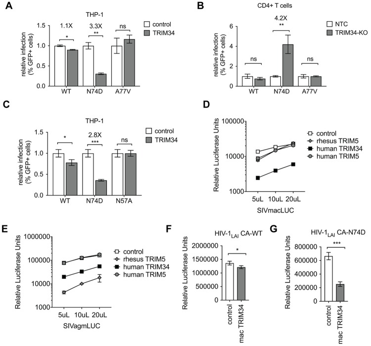
Fig 3. TRIM34 inhibits a range of primate lentiviruses independent of CPSF6.
A: THP-1 cells stably-overexpressing TRIM34 (gray bars) or control cells (white bars) were infected with WT, N74D or A77V HIV-1 and levels of infection assayed 2 days post-infection by flow cytometry. The relative infection is normalized to the average infection in the control cells for each virus. Data are represented as the mean+/- s.d. from triplicate infections. B: Primary, activated CD4+ T cells were electroporated with Cas9-RNP complexes targeting TRIM34 (gray bars: TRIM34-KO) or Non-Targeting Control crRNAs (white bars: NTC). 2 days later edited CD4+ T cell pools were infected with GFP reporter HIV viruses (WT, N74D or A77V) and infection levels assayed 2 days later by flow cytometry. Data is shown as infection levels relative to the Non-Targeting Control infections for each virus (relative infection). TRIM34-KO pool edited at 52% (ICE KO-score). Data are represented as the mean +/- s.d. from triplicate infections. C: THP-1 cells stably-overexpressing TRIM34 (gray bars) or control cells (white bars) were infected with WT, N74D or N57A HIV-1 and levels of infection assayed 2 days post-infection by flow cytometry. The relative infection is normalized to the average infection in the control cells for each virus. Data are represented as the mean+/- s.d. from triplicate infections. D and E: THP-1 cells were transduced with lentiviral vectors encoding rhesus TRIM5α (gray diamonds), human TRIM5α (gray circles), human TRIM34 (black squares) or a control vector (white squares). Each cell pool was infected with VSV-G pseudotyped SIVmacLUC (D) or SIVagmLUC (E) Luciferase reporter viruses at 3 viral doses as indicated. Levels of infectivity were assayed 2 days later by luciferase assay (RLU = Relative Luciferase Units). Data are represented as the mean +/- s.d. from triplicate infections. F and G: THP-1 cells stably-overexpressing rhesus macaque TRIM34 (mac TRIM34—gray bars) or control cells (white bars) were infected with WT (F) or N74D (G) HIV-1 and levels of infection assayed 2 days post-infection by luciferase assay. Data are represented as the mean+/- s.d. from triplicate infections. P values were determined using two-sided unpaired t-tests (ns = not significant, *P<0.05, **P<0.01, ***P<0.001, ****P<0.0001).

