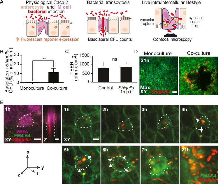
Fig 1. Establishment of a physiological in vitro model of S. flexneri M cell infections at high spatiotemporal resolution.
(A) Scheme of the FAE model and its applications with genetically encoded fluorescent reporters, translocation assays and confocal imaging. (B) S. flexneri translocates exclusively through M cell containing co-cultures at 1h post-infection (p.i.) and not through polarized monocultures, (n = 5, *P <0.01). (C) Co-culture barrier integrity, monitored by transepithelial electrical resistance (TEER) measurements in non-infected (control) and infected monolayers, is preserved at 1h p.i. (n = 5). (D) S. flexneri infection plaques form exclusively within M cell containing co-cultures (right panel) and not in polarized monocultures (left panel) at 21h p.i. (n = 5). Scale bar, 50 μm (E) Representative time-lapse of S. flexneri targeting a WGA-positive M cell at 1h p.i. before spreading to neighboring cells from 4 to 6h p.i. (arrows) and generating an infection plaque at 21h p.i.. Scale bar, 20 μm.

